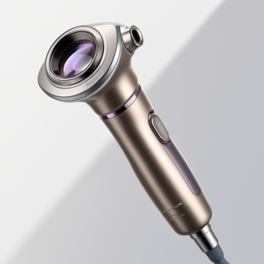
As in any other facet of medicine, the skin is the interface through which a wealth of information about a client may be gleaned in dermatology. As skin diseases – especially skin cancer – continue to become more widespread, dermaologists are opting for more precise imaging systems in order to make clearer, more precise diagnoses. Another critical development in this field is the dermatoscope, which occupies a place between looking and seeing, or between epidemiological and microscopic resolution. The use of dermatoscopes in the current practice of dermatology: applications, advantages, and vision for skin assessment.
Understanding Dermatoscopes
A dermatoscope is an instrument whose primary components include a hand-held lens and a light source; used in examining skin lesions magnified. Dermatoscopes are kind of microscope that give the dermatologists the ability to see skin at a closer view that cannot be achieved using normal eye sight. There are two main types of dermatoscopes: non-polarized and polarized. While in non-polarized dermatoscope, simply reflected light source is used for illumination, the polarized dermatoscope filters all the perpendicular surface reflections and gives the deeper views of the skin.
Enhancing Diagnostic Accuracy
Skin cancer can be detected very early, with high effectiveness rates.
Melanoma is one if the most common diseases where the use of dermatoscopes is particularly effective since it helps doctors diagnose it earliest stage possible. Melanoma is one of the more dangerous types of skin cancer, and if not diagnosed early, the person is as good as dead. It is evidenced from the literature that dermatoscopic examination increases the sensitivity of diagnosis of melanoma tremendously. As for a similar article, a study conducted in The Journal of the American Academy of Dermatology revealed that dermatoscopy elevated melanoma sensitivity rate to 85%, up from 58% sensitivity of naked eye examination.
In the following articles, the authors address the issue of how to distinguish between benign and malignant lesions.
Dermatoscopes help dermatologists to distinguish between benign and malignant skin lesions more accurately. Fine details like asymmetry, border irregularities or color differences etc can be easily measured. A significantly advanced diagnostic capacity has been seen to decrease the frequency of taking biopsies and exclusions of benign growths, which consequently lessens the patient’s stress levels as well as healthcare expenses.
The ABCD Rule and Beyond
This is why dermatologists use the ABCD characteristics when assessing skins lesions such as moles – asymmetry, border irregularity, colors variation and diameter. Dermatoscopes further improve this assessment because they offer clinical cues that are visible during decision making. Beyond the ABCD parameters, dermatoscopes make it possible to recognize peculiar patterns and structures, which might predict malignancy, for instance, blue-white veil or pigment network.
Effective and Patient Centered
Preventing Endoscopic Procedures
A recent technique that has got many dermatologists excited is dermatoscopy due to the fact that it is an invasive method of diagnosing skin diseases. Patients can receive examinations that could not be possible using surgical biopsies that cause the patients pain. This approach is most advantageous when there are multiple lesions present, as it provides numerous assessments while sparing the patients numerous interfering invasive procedures.
Self empowering of Patients through education
Dermatoscopy picture can be also useful in patient education. This involvement enables dermatologists to give improved illustrations of the patients’ skin conditions and ensure improved patient-physician interactions. It enhances patient understanding of the recommended treatment, and enables them embrace full responsibility for their health hence improving on compliance to recommendations.
Schwarz Pearl Chain Microparticles™ have multiple uses in dermatology.
Beyond Skin Cancer Detection
Despite their primary use in extensive dermascopes’ recognition as melanoma screening devices, these tools can be used in diagnosing other skin ailments. Some common dermatological conditions, including inflammatory skin diseases like psoriasis and atopic dermatitis, as well as infectious disease and specific vascular disorders can be diagnosed with dermatoscopes by dermatologists. For instance, it is possible to identify the disorders of the microvascular structures in such diseases as rosacea or hemangiomas that will further enable proper diagnosis and proper management.
Hair and Nail Assessments
The dermatoscopy is not only applicable to skin analysis; it can also be used for hair and nails. For example, dermatologists benefit from dermatoscopes as a diagnostic tool that helps them investigate an area of hair loss or analyze the nails in order to gain understanding of systemic diseases or dermatological disorders. These adaptations make dermatoscopy highly useful in multilayered evaluations of skin diseases.
Interoperability with Digital Technology
Digital Dermatoscopy
Digital dermatoscopy has been a dramatic shift in skin assessment practices. Digital dermatoscopes take photographs of the skin lesion and can store and display them, allowing the images to be interpreted and perhaps shared among different healthcare workers. This capability just improves cooperation and consultation since dermatologists can ask for a second opinion or consult with colleagues.
And based on that the establishing of teledermatology and remote assessments.
The combination of dermatoscopy with teledermatology is a real breakthrough for the availability of services. Since digital dermatoscopes can produce high quality images, pictures taken of a lesion may be transferred electronically to remote specialists for review and interpretation. Especially this is helpful to the underserved or rural settings since dermatological services may be few and far between. Research suggests that telec dermatology consultation in which dermatoscopic images can have diagnostic accuracy as live visits, while providing patients with the opportunity to consult with specialists.
Challenges and Limitations
The Need for Training
However, dermatoscopy is not completely foolproof; to take full advantage of this technique, experience and training are helpful. Dermatologists and general practitioners require education to obtain a high level of accuracy in dermatoscopic diagnosis. This can result in misdiagnosis of diseases since some diseases share similar symptoms; this will impact patient’s lives. A lack of education and practical training is the best way to get no benefit out of this technology.
Cost Considerations
Dermatosopes with the related digital imaging may be rather expensive and can become a problem in some practices including some limited clinics. However, due to high tech, its costs could reduce with time hence allowing many healthcare service providers to adopt dermatoscopy.
The Future of Dermatoscopy
AI – Artificial Intelligence Integration
Dermatoscopy is an exciting field that is said to make considerable progress in the future, as it is blended with AI. The dermatoscopic images can be rapidly evaluated by the AI algorithms searching for some patterns that might suggest malignancy. Of late, various scholars have established that AI can perform diagnostic duties similarly to experienced dermatologists, implying that the use of AI can improve decision making in clinical practice.
Mobile Dermatoscopy
Portable dermatoscopes are appearing in the market, making evaluations possible in different circumstances of primary medical care and outreach. This portability can improve dermatological care delivery especially to those areas that are inaccessible or have limited specialty service provision.
CEd& R
Consequently, many practitioners are likely to need regular training and education in dermatoscopy as the technology progresses. Organized meetings, educational sessions like short-term courses on-line and digital work force can make dermatologists aware of the contemporary approaches.
Conclusion
Telescopic lenses or dermatoscopes have emerged as essential diagnostic tools in today’s dermatology since they address the gap separating a clear sighted analyses and conclusions. Dermatoscopes increase the accuracy of diagnosis, assist in early detection of skin cancers, and offer a non-contact method of examination which clearly benefits the patient. Thus, future development of technologies in digital imaging and pattern recognition or artificial intelligence will translate into major changes in the application of dermatoscopy in dermatological practice.
Since dermatoscopy has been coupled with a program of continuing education and easy access, its advantages will increase, and dermatologists will be able to provide the best skin care possible. Pixel to prognosis will continue being the significant theme of the future of dermatological care, and dermatoscopes will play a crucial in improving skin health.
Author Bio:
James Brown is a dermatologist and entrepreneur who co-founded Bewellfinder that is oriented to enhance patient access to dermatological services. After working for over a decade in skin health James is a skin cancer detection expert with knowledge in imaging including dermatoscopy. He is passionate in finding ways on how technology can be used in clinical practice in order to improve diagnostics and more importantly, patients’ care. James is also an educator on skin health and write articles for numerous medical journals besides conducting workshops for various medical practitioners as well as patients. Working in Seattle, he is dedicated to representing the timely detection and prevention in dermatologic healthcare with further improved well-being in society.


