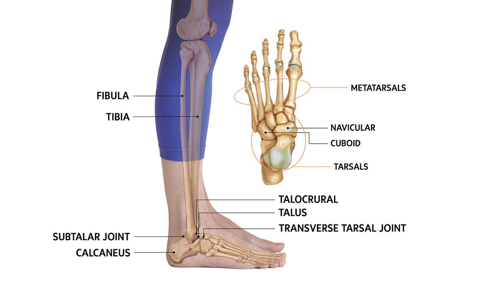
The ankle-joint fracture is often injured in adults. This joint is concerned with movements in the anteroposterior direction and during plantarflexion and dorsiflexion. While walking, the foot is fixed on the ground and any rotational violence tends to deviate the ankle from the normal anteroposterior movements. These abnormally directed forces are external rotation, internal rotation, abduction, and adduction which tend to produce undue strain on the joint. The external rotation tends to produce bony injury whereas internal rotation usually produces ligamentous lesion. Injuries can manifest in the form of sprain, rupture of the ligament, fracture, and dislocation of the joint. The nature of violence which produces these various lesions differs only in the severity of the force and with the increase of its character the seriousness of the lesion increases likewise.
Ankle sprain
Mechanism of injury
Itis the lateral ligament which is commonly affected because of an inversion injury. The sudden placing of the foot on an uneven surface may produce inversion rotation and the lateral ligament is stretched. Most of the component fibres of the ligament are intact and only a few fibres may be torn.
Diagnosis
Clinical examination: There are pain, swelling, ecchymosis on the outer aspect of the joint. Tenderness is elicited below the tip of the lateral malleolus.
X-ray: X-ray should be taken to exclude any fracture of the lateral malleolus. When a rupture of the lateral ligament is suspected stress X-ray of the ankle-joint is taken under general or local anesthesia. The lateral tilt of the talus is seen on inversion strain. When the lateral ligament is ruptured, for clarity the normal joint may be x-rayed for comparison.
Treatment
Adhesive strapping is applied to extend from below the knee up to the metatarsal heads. The foot is placed in an everted position. Thepatient is encouraged to walk from the beginning. Strapping is removed after two weeks.
Rupture of the lateral ligament
Mechanism of injury
This is produced in the same way as the inversion injury. The nature of violence tends to be more severe than the previous one.
Diagnosis
There is usually more swelling and more bruising than the sprain.
X-ray: X-ray is taken under general or local anaesthesia after applying inversion strain to observe any gaping on the outer aspect of the joint space with a lateral tilt of the talus.
Treatment
Treatment is based on whether the case is an early one or of the chronic variety.
- Early-stage: Thisis a serious injury and chronic instability of the joint may be produced if the ruptured ligament does not unite.
- Immobilization: A short-leg walking plaster extending from below the knee to the metatarsal heads is applied. The foot is maintained in a state of eversion. The plaster is removed usually after 2 months.
- Immediate repair: When evidence of the ligamentous tear is present, immediate operative repair and immobilization by plaster cast can be done. Operative repair requires some ortho implant.
- Chronic cases: The patient complains about the ankle giving way during the time of walking. This happens because of the absence of any support on the lateral aspect of the joint when exposed to inversion strain.
Shoe appliance: Patient is advised to wear a shoe with the outer border raised and the inner side depressed.
Operation: New lateral ligament is constructed by tenodesis using the peroneus brevis tendon.


